

立即搜索


产品介绍
Source
Chinese Hamster Ovary cell line
Description
Human Fibronectin Fragment
Accession # P027501
Predicted molecular mass
62.6 kDa
Specification:
Appearance
White powder, Colorless clear liquid after reconstitution
Purity
≥95%, by SDS-PAGE (under reducing (R) & Non-reducing conditions, visualized by Coomassie staining)
Endotoxin
≤10 EU/mg by the LAL method
Activity
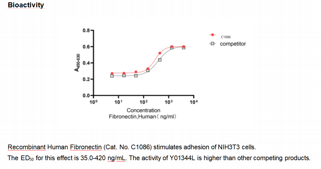
Measured by the ability of the immobilized protein to support the adhesion of B16‑F1 mouse melanoma cells. The ED50 for this effect is 35.0-420 ng/mL.
Formulation
Lyophilized from a 0.22 μm-filtered solution containing PBS, 5% mannitol and 0.01% Tween 80, pH 7.4
Size
50 μg/vial, 1 mg/vial
Handling and Storage:
Reconstitution
It is recommended to redissolve in sterile deionized water.
Shipping
The product is shipped with blue ice.
Storage & Stability
24 months at 2°C to 8°C in lyophilized state
7-10 days at 2°C to 8°C under sterile conditions after reconstitution
6 months at -20°C to -80°C under sterile conditions after reconstitution
Use a manual defrost freezer and avoid repeated freeze-thaw cycles.
 |
Background:
This protein is a secreted, glycosylated cytokine composed of four alpha helical bundles. The encoded protein is mainly synthesized in the kidney, secreted into the blood plasma, and binds to the erythropoietin receptor to promote red blood cell production, or erythropoiesis, in the bone marrow. Expression of this gene is upregulated under hypoxic conditions, in turn leading to increased erythropoiesis and enhanced oxygen-carrying capacity of the blood. Expression of this gene has also been observed in brain and in the eye, and elevated expression levels have been observed in diabetic retinopathy and ocular hypertension. Recombinant forms of the encoded protein exhibit neuroprotective activity against a variety of potential brain injuries, as well as antiapoptotic functions in several tissue types, and have been used in the treatment of anemia and to enhance the efficacy of cancer therapies. [provided by RefSeq, Aug 2017]
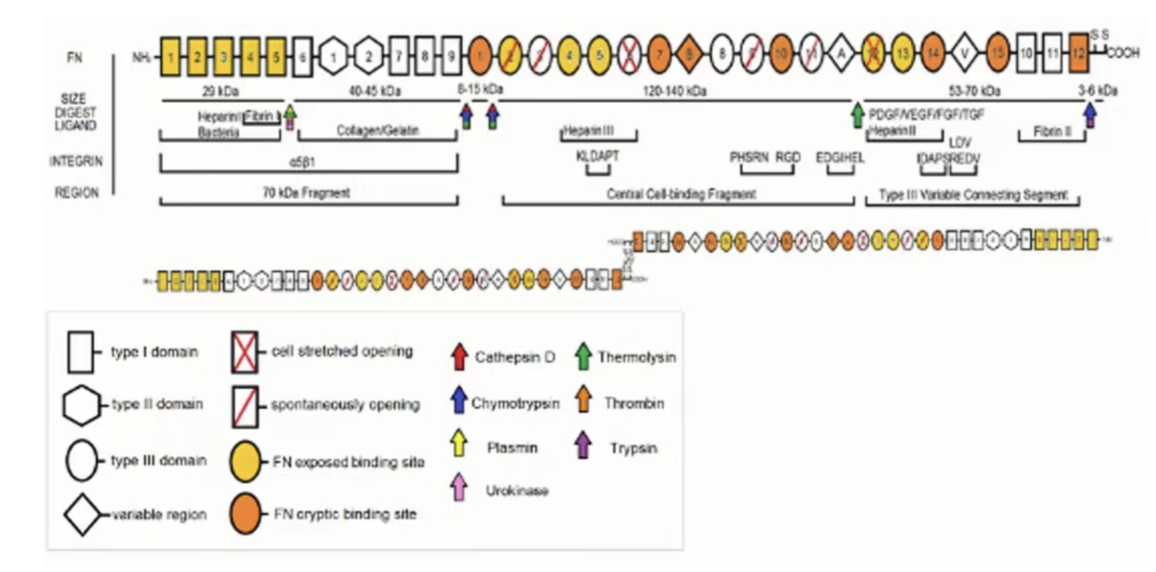
Fibronectin (FN) is a glycoprotein whose size ranges from 230 to 270 kDa and usually exists as a dimer, covalently linked by a pair of disulfide bonds at the C-termini. Each monomer consists of three repeating units: 12 Type I, 2 Type II, and 15-17 Type III domains which combined account for 90% of the FN sequence. FN circulating in the blood and produced by cells provides the basis of the extracellular matrix (ECM) formed in healing acute wounds. The time-dependent deposition of FN by macrophages, its synthesis by fibroblasts and myofibroblasts, and later degradation in the remodeled granulation tissue are a prerequisite for the successful healing of wounds.
 |
图 FN结构域
[1] Dalton CJ, et al. 2021. Cells. 10(9):2443.
[2] Kanta J, et al. 2020. Vasc Med. 25(6):588-597.
仅供科研或生产使用,不可直接应用于人体。
规格/储存条件/有效期
| 规格 | 1mg |
| 储存条件 | -20℃ |
| 有效期 | 24个月 |
COA下载
